Introduction
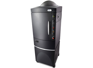
The PerkinElmer IVIS® SpectrumCT preclinical in vivo imaging system is an integrative platform that combines the full suite of IVIS optical features including Spectral Unmixing, 2D and 3D quantitative bioluminescence and fluorescence with fast and low dose CT imaging, ideal for longitudinal studies.
The system provides researchers with greater insights into complex biological systems by enabling simultaneous molecular and anatomical non-invasive imaging in animal models.
Fast imaging and the ability to image multiple animals offers the throughput required to scan large cohorts of animals quickly and draw sound conclusions from your experimental data.
The system is equipped with an isoflurane anesthesia system, low-autofluorescence optics and a cooled CCD camera (‑90°C) in addition to the X-ray source.
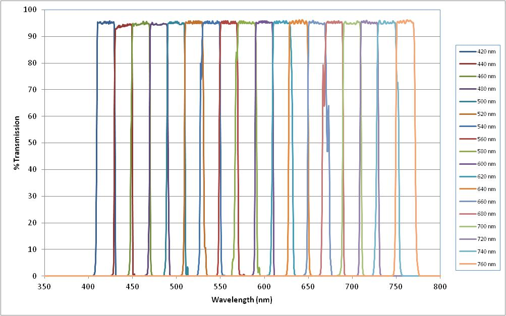
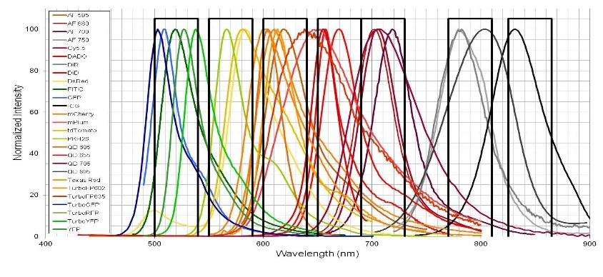
The IVIS® SpectrumCT is equipped with 28filters (10 excitation filters and 18 emission filters) to image fluorescent sources that emit from green to near-infrared. The X-ray module employs automated image integration to overlay with bioluminescence, fluorescence and photography.
The system is located at the Gutwirth Bldg. at the Technion main campus.
Available fluorescence filters
| 10 Excitation filters
/30 nm Bandwidth |
18 Emission filters
/20 nm Bandwidth |
|---|---|
| 430 | 500 |
| 465 | 520 |
| 500 | 540 |
| 535 | 560 |
| 570 | 580 |
| 605 | 600 |
| 640 | 620 |
| 675 | 640 |
| 710 | 660 |
| 745 | 680 |
| 700 | |
| 720 | |
| 740 | |
| 760 | |
| 780 | |
| 800 | |
| 820 | |
| 840 |
Applications
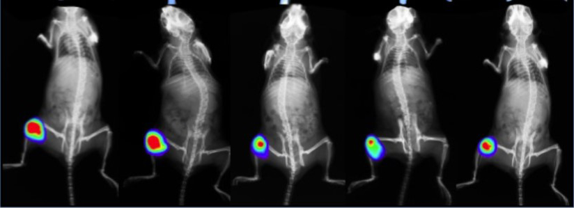
- Quantitative longitudinal measurement of tumor development, metastasis, angiogenesis, and response to therapy
- Trafficking of cellular and other biological therapies (e.g., immune cell, stem cell, or viral therapeutic platforms or antibodies, peptides, metabolites, etc.)
- Monitoring of host immune cell responses
- Distribution, quantification, and kinetic analyses of gene expression or enzymatic activity in vivo
Key features include:
Integrated optical and microCT technology
3D optical tomography for fluorescence and bioluminescence
High sensitive detection technology ideal for:
Bioluminescence
Multispectral fluorescence and spectral unmixing
Cerenkov imaging for optical radiotracer imaging
Low dose and ultra fast microCT
DyCE™ dynamic enhanced imaging for real time distribution studies of both fluorochromes and PET tracers ideal for PK/PD app
Analysis
The IVIS Spectrum CT Living Image software allows absolute quantification (photons/sec/cmsq/sr) of bioluminescence and fluorescence signal, for in vivo, ex vivo and in vitro studies.
The software supports accurate autofluorescence removal, spectral unmixing, fluorophore quantitation, automatic ROI creation, background subtraction and scaling.
The software is available IVIS Spectrum -CT work station.
New users
Please contact Dr Galit Saar, tel. +972-73-3785347 to coordinate a meeting.
Reading materials prior to the first instruction session: IVIS Spectrum-CT Basics and Living Image User manual
The system can be reserved in the BookItLab online ordering system and is located in the Gutwirth Bldg. at the Technion main campus.