Electron Microscopy

Scanning and Transmission Electron Microscopy
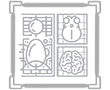
Electron microscopy is a powerful magnification tool, designed to detect fine details at high magnifications (sub-nm).
A wide range of samples can be observed using scanning and transmission electron microscope, including cells, cell organelles, viruses, cell infection assays, tissues and tissue slices, bacteria and bacterial interactions, fungi and many more. Gold-labeled secondary antibodies can be used to visualize antigens located on the surface or inside the sample.
We have an in-house Talos L120C transmission electron microscope at the faculty and we offer sample preparation and acquisition of scanning and transmission electron microscopy data of:
- Classic rendering of the surface and content of the sample.
- Immuno-gold labeling of surface-located antigens.
- Resin embedding and ultramicrotomy
- Immune-gold labeling of thin sections
- Negative staining
Please contact Lihi Shaulov, tel. 073-378-1125 for more information and to coordinate a meeting to discuss the possibilities.
Academic Head: Assoc. Prof. Tom Schultheiss.