Sample Preparation for SEM
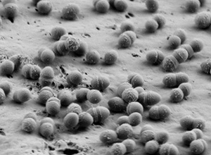
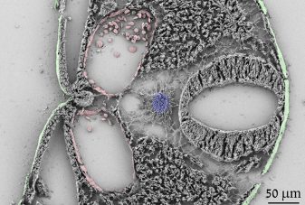
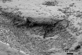
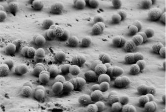
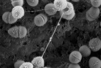
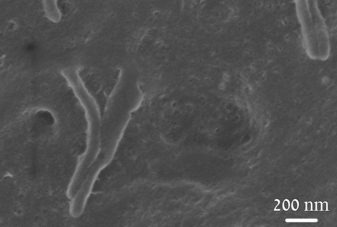
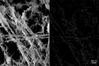
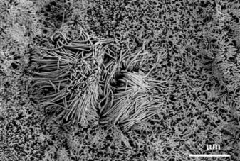
High resolution Scanning Electron Microscopy (SEM) allows visualization of microstructural characteristics of specimens at the nanometer to micrometer scale. We offer biological sample preparation for high resolution SEM to visualize the surface topology. A wide range of samples can be observed including cells, cell organelles, viruses, cell infection assays, bacteria and bacterial interactions, fungi and many more. Gold-labelled secondary antibodies can be used to visualize antigens located on the surface of the sample.


