Gallery
Chick Embryo Slice
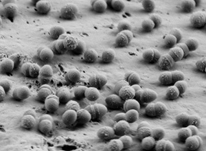
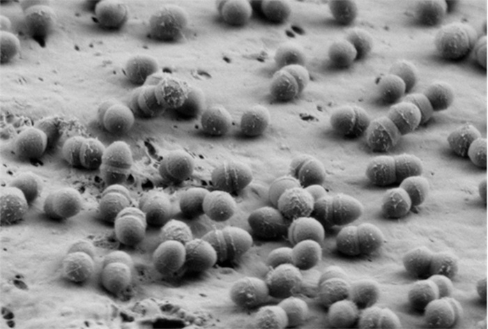
Bacteria
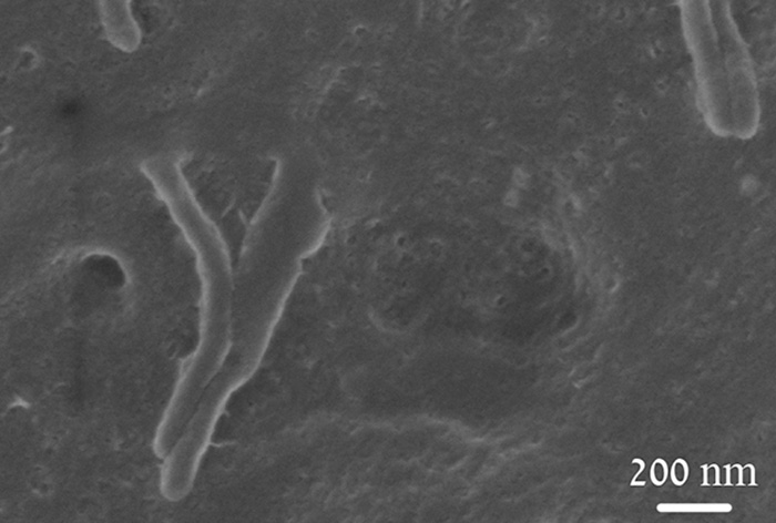
Bacteria

Bacteria
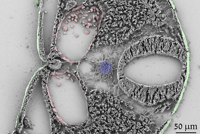
Fibroblast
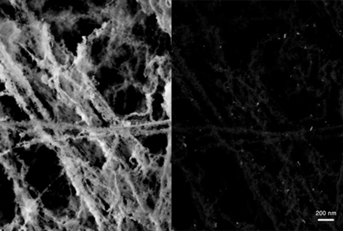
Fibroblast ESM and Fibronectin Immunogold
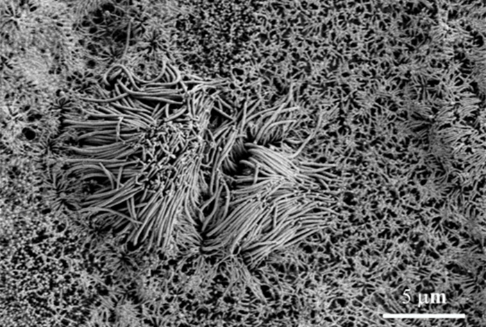
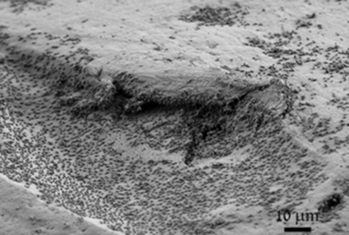
Lung Cilia
