Live Cell Imaging

We provide a range of options for both short-term and long-term live cell imaging:
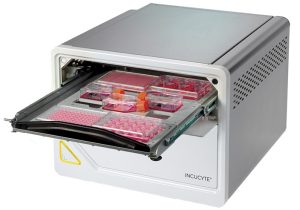
High Throughput Widefield Imaging
- Incucyte SX5: This system can handle up to six multiwell plastic bottom dishes. It supports imaging in both phase and fluorescence (excluding DAPI) with objectives of x4, x10, and x20 long working distance.
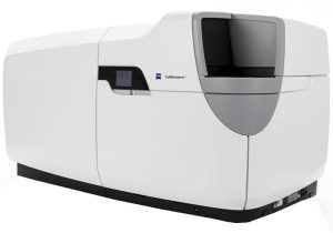
High Content Widefield Imaging
- Celldiscoverer 7: This versatile system supports transmitted light or fluorescence imaging across all channels. It accommodates a single multiwell plate, 3.5 or 6 cm petri dish, or three chamber slides. The Celldiscoverer 7 offers long working distance for plastic bottom dishes and high-resolution air or water immersion objectives, with magnifications ranging from x2.5 to x100.
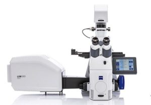
Laser Scanning Confocal Imaging
- LSM900 Inverted: Equipped with an on-stage incubator, this system supports confocal imaging with Airyscan for one 35 mm petri dish or one chamber slide.
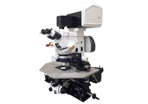
- LSM880 and LSM900 Upright Systems: These systems feature water dipping objectives for petri dishes, providing confocal imaging with Airyscan. Note that these systems do not include an incubation option.
Spinning Disk Confocal Imaging
- Olympus IXplore-Spin: This spinning disk confocal system offers faster and gentler imaging compared to scanning confocal systems. It includes an on-stage incubator that can accommodate a multiwell dish, petri dish, or chamber slide.
Please contact Maya Holdengreber, tel. 073-378-1106, to coordinate a meeting.