
Pratibha Ahirwal

Matteo Ghiringhelli

Matteo Ghiringhelli
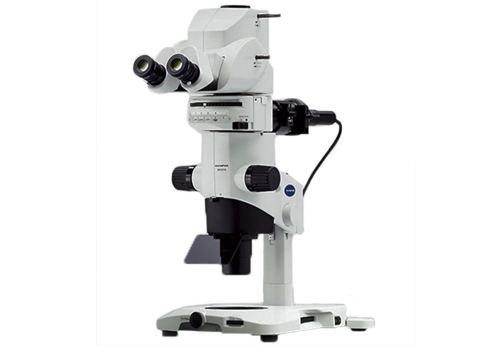
The Olympus MVX10M research macro zoom microscope images brightfield and fluorescent specimens, such as Drosophila larvae, organs of small animals, and more.
DP73 is a 17.28 megapixel color CCD (2 megapixels with pixel shifting), 12bit
1600×1200 pixel images at 15 fps, 800×600 at 27fps
| Objective | Magnification/NA | Working distance (mm) | Total magnification |
|---|---|---|---|
| MVPLAPO | x1/0.25 | 65 | 6.3x-63x |
| MVPLAPO | x2/0.5 | 20 | 12.5x-125x |
The x2 objective has a correction collar for correction of aberration produced by water or plastic container.
Fluorescence: X-cite 120PC –Q-metal halide lamp
Incident Illumination: Dual Olympus tungsten-halogen dichroic reflector
Transmitted Illumination Base: LED four position turret (darkfield, brightfield, oblique).
| Filter | Excitation Filter | Dichroic | Emission Filter | Fluorophore Family |
|---|---|---|---|---|
| U-XL49000 | AT350/50 | T400LP | ET460/50 | DAPI |
| U-XL49002 | ET470/40 | T495LPXR | ET525/50 | Cy2,EGFP |
| U-XL49004 | ET545/25 | T565LPXR | ET605/70 | CY3,RFP |
Acquisition is performed with the Olympus proprietary Cell Sens Dimension software.
Output files can be opened with FIJI/ImageJ using the Olympus viewer plugin or exported to TIFF.
Please contact Maya Holdengreber, tel. 073-3781107, to coordinate a meeting.
The Olympus Stereoscope can be reserved in the BookItLab online reservation system.
