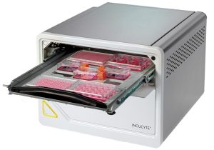
Other Inverted Incubated Widefield Systems
Zeiss Celldiscoverer 7

Introduction
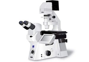
Celldiscoverer 7 is a fully integrated, automated high-content screening live cell inverted widefield imaging system for cell cultures, tissue sections or small model organisms with optional controlled environmental conditions including CO2, O2, N2, heating and cooling. It features built-in automation for easy setup of complex experiments. High-resolution experiments can utilize automatic water immersion supply and removal which suits even long-term experiments.
Hardware
Cameras
The system has two monochrome cameras:
LED illumination and filter sets
| LED combinations (nm) | Filter set | Beamsplitter | Emission filter |
|---|---|---|---|
| 385 / 470 / 567 / 625 / IR-TL | 90HE | 405+493+575+653 | 425/30 + 514/30 + 592/25 + 709/100 |
| 420 / 520 / 590 / IR-TL | 91HE | 450+538+610 | 467/24 + 555/25 + 687/145 |
| 385 / 470 / 590 / IR-TL | 92HE | 405+493+610 | 425/30 + 524/50 + 688/145 |
Objective lenses with temperature control
| Objective | Magnification | NA | Vessel thickness | Effective penetration | Working distance (mm) | Immersion |
|---|---|---|---|---|---|---|
| ZEISS Plan-APOCHROMAT 5× / 0.35 |
x5 | 0.35 | Phase 1 | up to 1.2mm | air | |
| ZEISS Plan-APOCHROMAT 20× / 0.7 |
x20 | 0.7 | Phase 1 | 0.13-1.2mm with autocorrection |
1.33mm @ 0.17mm 0.4mm @ 1mm |
air |
| ZEISS Plan-APOCHROMAT 20× / 0.95 |
x20 | 0.95 | Phase 2 | 0.13-0.21mm with autocorrection |
0.4mm @ 0.17mm | air |
| ZEISS Plan-APOCHROMAT 50× / 1.2 W |
x50 | 1.2 | Phase 2 & DICII | 0.13-0.21mm with autocorrection |
0.4mm @ 0.17mm | water autoimmersion |
Sample mounting
- One multiwell dish
- One 35mm or 60mm petri dish
- Three 76x26mm slides
- Two slides/Lab-TekTM chambers 57×26 mm
- Insert plate for perfusion with POC-R2
Acquisition and Analysis
Acquisition is performed with the Zen software which includes modules for automated quantification of fluorescence, cell confluence by transmitted light including wound healing and spot detection inside cell nuclei. For basic visualization and processing, you can download the free ZEN lite edition software. Offline full versions are available for analysis at CT Computer and Computer IMARIS software. Output files can be opened by Imaris and ImageJ/FIJI for analysis.
New users
Please contact Maya Holdengreber, tel. 073-378-1106, to coordinate a meeting.
Tips for immunofluorescence experiments.
Please prepare controls before starting a new model.
Reservations
For live widefield microscopy, the Celldiscoverer 7 can be reserved through the BookItLab online reservation system.


