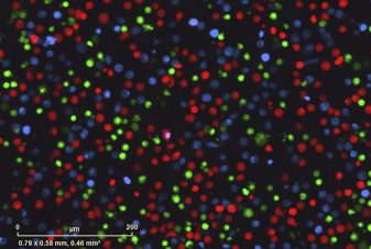
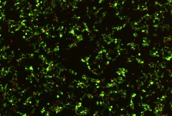
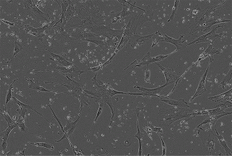
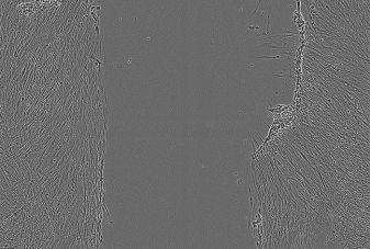
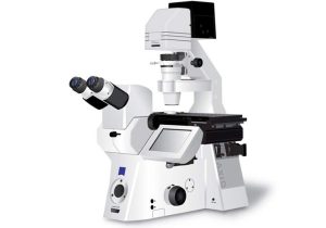
Other Inverted Incubated Widefield Systems
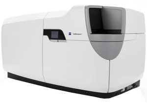
Incucyte SX5

Introduction
The Sartorius Incucyte SX5 System is a high throughput, continuous live cell imager.
It enables kinetic transmitted (phase) and fluorescence imaging of long term live cell experiments inside the stable environment of an incubator without sacrificing cells to obtain data from each timepoint. The multiwell dish remains stationary and the microscope moves to image.
Hardware
Camera
Basler acA 1920-155um – Basler ace, with the Sony IMX174 CMOS sensor, 1920 px x 1200 px, 12bit.
Objectives
| Objective | Magnification/NA | Pixel size |
|---|---|---|
| Nikon Plan Apo λ | x4/0.20 | 2.82 μm |
| Nikon Plan Fluor | x10/0.30 | 1.24 μm |
| Nikon S Plan Fluor | x20/0.45 | 0.62 μm |
All objectives have long working distance; many plastic off-the-shelf vessels can be used.
Channels
| Channel | Excitation | Emission | |
| Green Orange NIR |
Green Orange NIR |
453-485 nm 546-568 nm 648-674 nm |
494-533 nm 576-639 nm 685-756 nm |
| Green Red |
Green Red |
441-481 nm 567-607 nm |
503-544 nm 622-704 nm |
| Metabolism | ATP | 473-498 nm 524-550 nm |
565-591 nm |
| Orange NIR |
524-550 nm 648-674 nm |
565-591 nm 685-756 nm |
Acquisition
The proprietary software performs both acquisition and analysis. The software can be installed on any computer in the Technion and the experiment can be monitored in real time. The acquired images can be exported to JPEG, TIFF, TIFF series, MPEG4, AVI and the graphs can be exported as excel files or graphs.
Applications
Cell Counting and Proliferation
Viability
Apoptosis
Cytotoxicity
Tumor Spheroid
Cell Cycle
ATP Metabolism
Mitochondrial Membrane Potential
Immune Cell Killing
Antibody Internalization
Phagocytosis
NETosis
Neuronal Activity
Angiogenesis
Live-Cell Immunocytochemistry
Scratch Wound Migration
Invasion
Chemotaxis
Immune Cell Activation
New users
Please contact Maya Holdengreber, tel. 073-3781106, to coordinate a meeting.
Reservations
The Incucyte SX5 can be reserved through our BookItLab online reservation system.


