Related Technologies
Anatomy & Relaxometry
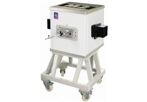
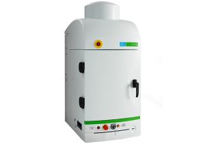
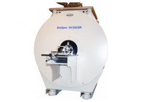
To monitor physiological changes and pathological development in animals, our facility offers various noninvasive in vivo imaging systems: ASPECT 1T MRI, Bruker BioSpec 9.4T MRI and Vevo 3100 Ultrasound. The ASPECT 1T MRI can provide whole animal anatomical images. Specifically, one can evaluate and monitor the anatomy of organs of interest such as limbs, spine, kidney layers, and lungs. To image smaller structures such as the pancreas and ovaries or specific brain structures (hypothalamus, ventricles, corpus callosum etc.) we recommend the Bruker BioSpec 9.4T MRI, which has higher sensitivity and resolution.
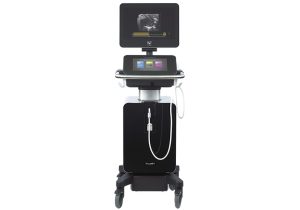
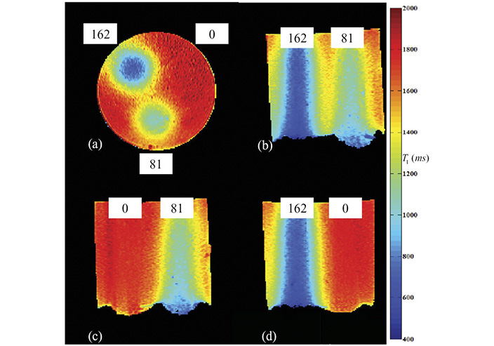
Another method that can be implemented by MR imaging is relaxometry. T1, T2 and T2* relaxometry maps of areas of interest can provide quantitative tissue characterization. For example, using T2 relaxometry one can differentiate between brain lesions and cancerous tissue. In addition, T1 or T2 relaxometry can shed light on tissue permeability using contrast agents. The Vevo 3100 Ultrasound is mainly used for abdominal organs (kidneys, intestines, pancreas, ovaries etc.), embryo development and 2D heart functional imaging.
Please contact Galit Saar tel. 073-378-5347 to coordinate a meeting.